AI for Radiology.
Accurate. Fast. Comprehensive.
PathoFinder measures your X-rays with models built in-house. A complete, annotated report is ready in seconds, and your radiologist reviews and approves it.
- 99.6%
- Agreement with expert radiologists, up to
- 3–6s
- To a complete, annotated report
- 30+
- Measurements in a single study, up to
One platform, three ways to measure
Each product reads a standard X-ray and returns the measurements your clinicians use, the way the best surgeons want them.
Alignment
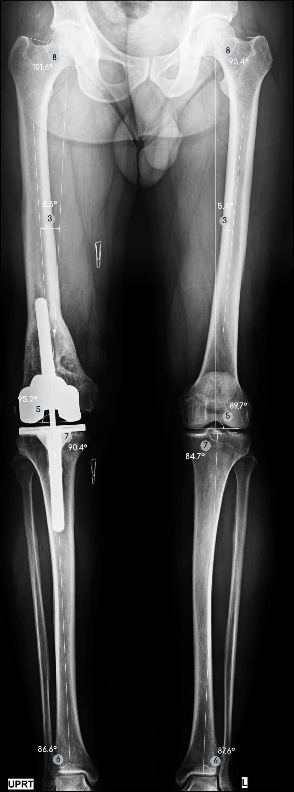
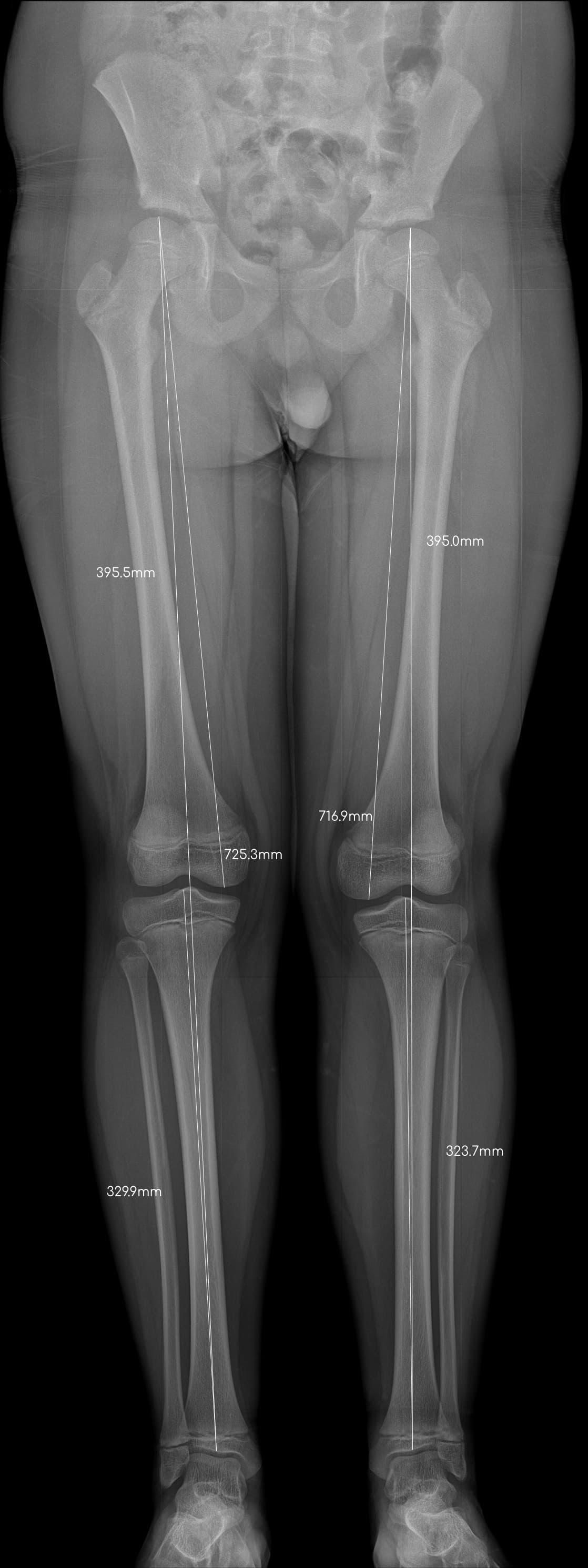
The full-length alignment report, measured the way the best knee surgeons want.
- Accurate
- 99.6%
- Agreement with expert radiologists
- Fast
- 3s
- Upload to full report. Manual: 10-15 min
- Comprehensive
- 15
- 8 angles, 6 lengths and CPAK class
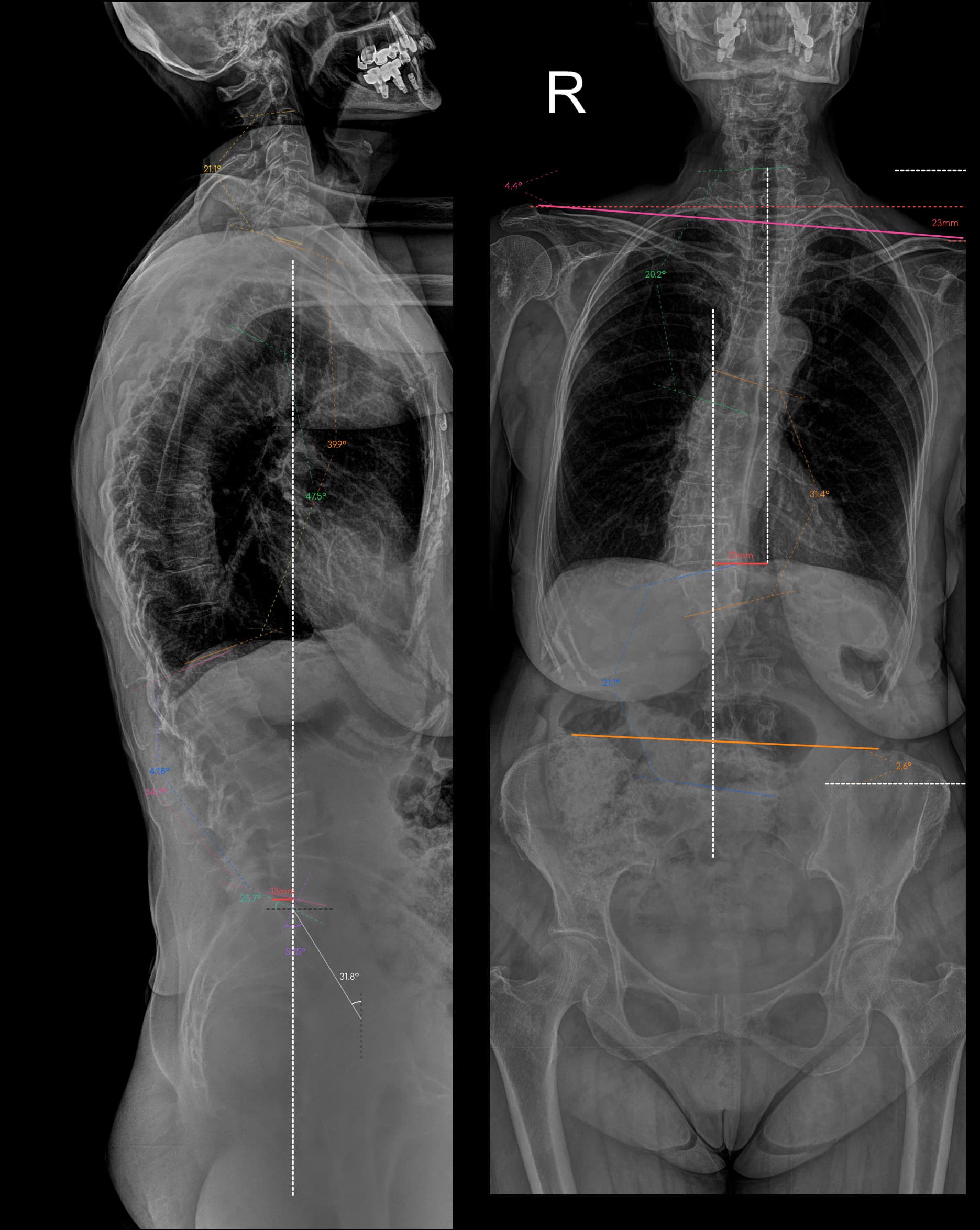
Full Spine
Complete coronal and sagittal spinal alignment, measured the way the best spine surgeons want.
- Accurate
- 99.3%
- Agreement with expert radiologists
- Fast
- 6s
- AP and lateral to report. Manual: 10-20 min
- Comprehensive
- 30+
- Coronal and sagittal parameters
Bone Age
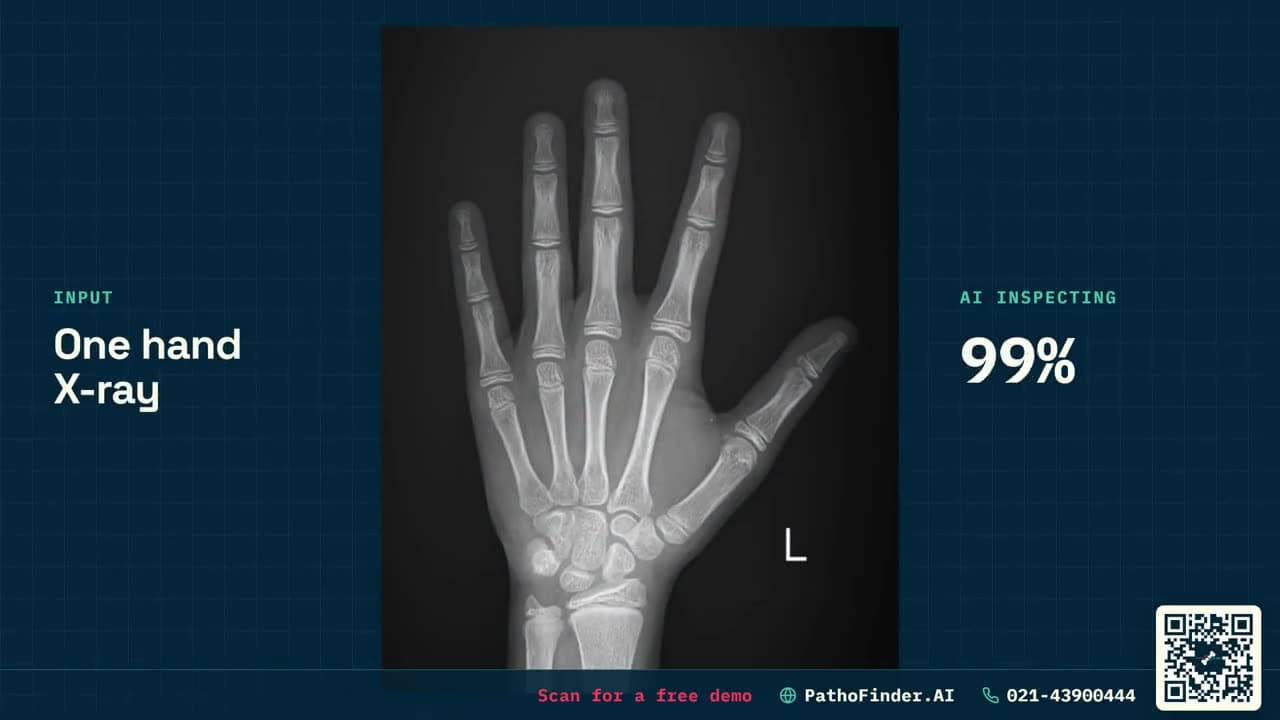
Skeletal maturity from a single hand X-ray, with the evidence behind every reading.
- Accurate
- 2.6mo
- Mean absolute error
- Fast
- 4s
- Upload to full report
- Comprehensive
- 7
- Regions per hand, SDS and growth remaining
Stitch
Joins separate X-rays into one full-length limb or spine image. Use it on its own or with Alignment and Full Spine.
Some of our products
A selection of the ML models we have built so far. We keep developing new ones, so the range changes often. Browse by body region or search by name.
Precision tools for radiology
A comprehensive platform designed to automate anatomic landmark localization for ankle, hip, knee, spine, alignment radiographs, and other imaging modalities.
Landmark localization
Automated identification of key anatomic reference points with sub-millimeter precision for orthopedic analysis.
DICOM support
Native support for DICOM formats ensures seamless integration with PACS and hospital information systems.
High-speed processing
Get results in under 1.5 seconds, allowing for real-time clinical decision support during patient visits.
Clinical accuracy
Our models are validated against expert-annotated radiographs and keep improving with every release.
Secure architecture
Enterprise-grade security ensuring patient data privacy and compliance with local healthcare regulations.
Standardized reports
Generate consistent, data-driven reports with annotated radiographs and precise measurement data.
How PathoFinder works
From automatic image intake to a finished report, in four simple steps.
Images arrive automatically
PathoFinder is installed locally at your site. Through our integration with your scanner or PACS, images enter the system on their own, with no manual upload.
AI processing
Our deep learning models localize anatomic landmarks and identify regions of interest.
Review results
Examine AI-generated coordinates, confidence scores, and landmark accuracy.
Publish reports
Generate comprehensive radiology reports and share findings securely with your clinical team.
The same two reports for every finder
Whatever the study, doctors get one familiar layout: the image with the findings marked, and the numbers in words.
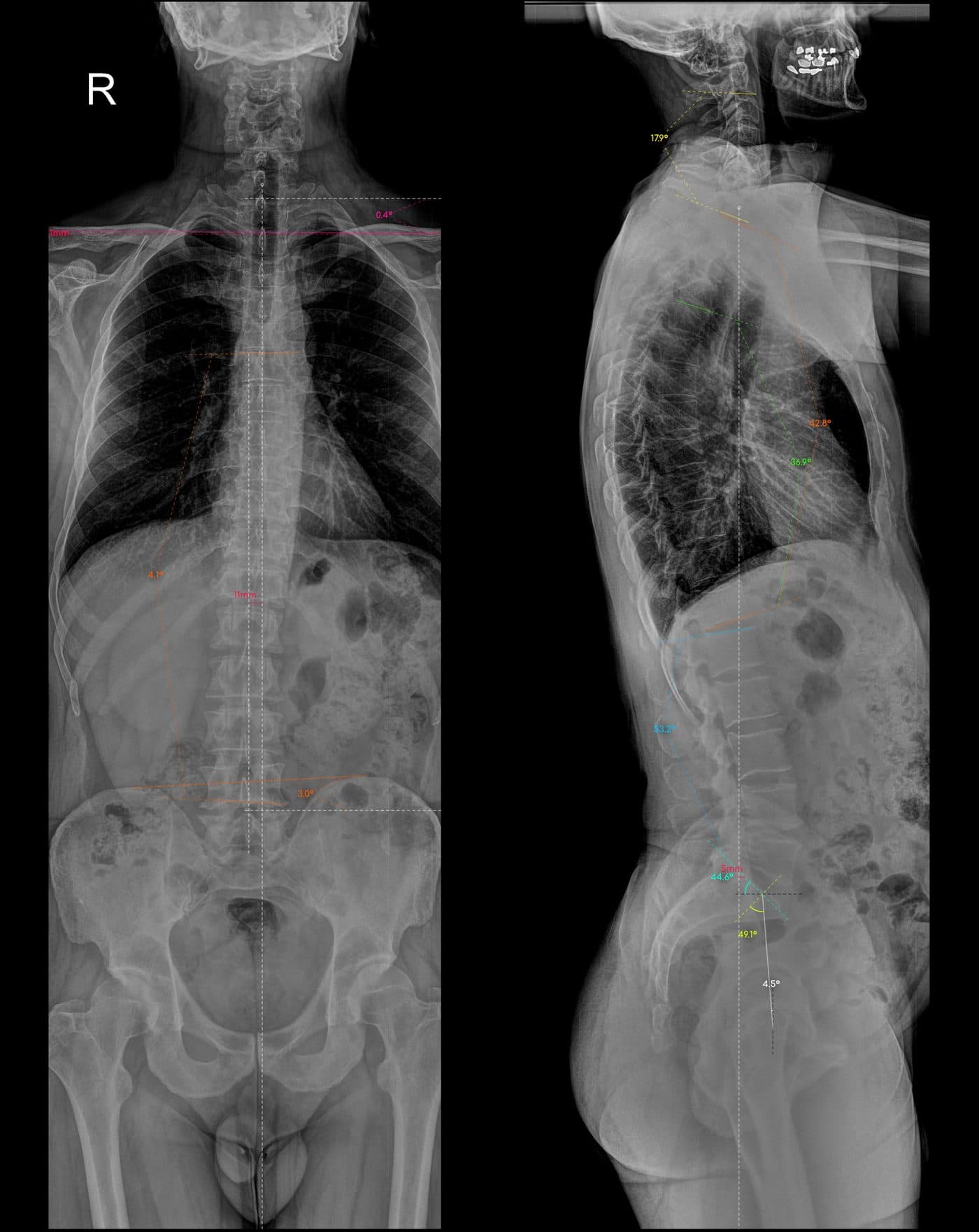
| Category | Parameters | Value |
|---|---|---|
| Curve I (Major) | Cobb | 4.1° |
| Region | Lumbar | |
| Convexity | Right | |
| UEV | T6 | |
| LEV | L4 | |
| Apex | L2 | |
| AVT | 3.0 mm | |
| Coronal balance | CVA | 11.0 mm |
| Direction | Left | |
| Pelvis | Pelvic obliquity | 3.0° |
| Side high | Left | |
| Shoulders | Acromion height difference | 1.1 mm |
| Side high | Right | |
| Clavicle angle | 0.4° | |
| Clavicle side high | Right | |
| Stable vertebra | Level | L5 |
| Category | Parameters | Value |
|---|---|---|
| Thoracic Alignment | T1-T12 kyphosis | 42.8° |
| T4-T12 kyphosis | 36.9° | |
| T5-T12 kyphosis | 27.8° | |
| T4-T9 kyphosis | 36.8° | |
| Lumbar Alignment | L1-S1 Lordosis | 53.1° |
| Cervical Alignment | C2-C7 Lordosis | 17.9° |
| SVA (C2-C7) | 12 mm anterior | |
| Pelvis | PI | 49.0° |
| PT | 4.5° | |
| SS | 44.6° | |
| Global Sagittal Alignment | SVA (C7-S1) | 5 mm posterior |
| C7 translation ratio | -2.24 | |
| T9SPi | 8.7° | |
| SSA | 136.5° | |
| Spinal tilt | 92.0° |